
Understanding the intricate head and neck anatomy is crucial, aided by resources like PDF atlases and comprehensive textbooks, such as Netter’s and Gray’s, for students.
These materials offer detailed illustrations and explanations, simplifying complex structures for effective learning and clinical application within dental and medical fields.
The study of this region benefits from accessible PDF guides, enhancing comprehension of the head and neck’s complex anatomical relationships.
Importance of Studying Head and Neck Anatomy
A thorough grasp of head and neck anatomy is paramount for numerous healthcare professionals, extending beyond medical doctors and dentists to encompass surgeons, radiologists, and speech therapists. PDF resources, alongside traditional textbooks, facilitate this essential knowledge acquisition.
This region’s complexity, housing vital structures like cranial nerves, major blood vessels, and intricate muscle groups, demands detailed study. Accurate anatomical understanding directly impacts diagnostic accuracy, surgical planning, and effective treatment strategies. For instance, comprehending the spatial relationships of structures is critical during surgical procedures to avoid iatrogenic injury.
Furthermore, the availability of head and neck anatomy PDF atlases and guides allows for convenient, self-paced learning and quick reference. Mastery of this anatomy is not merely academic; it’s fundamental to providing safe and effective patient care, ensuring optimal outcomes in a wide range of clinical scenarios.
Scope of Head and Neck Anatomy
The scope of head and neck anatomy is remarkably broad, encompassing a diverse array of structures from the skull and facial bones to the intricate network of muscles, nerves, and vessels. Accessible PDF resources, alongside comprehensive textbooks like Netter’s, aid in navigating this complexity.
It extends beyond mere bone structure, delving into the detailed arrangement of the scalp layers, the calvarium’s foramina, and the fascial compartments of the neck. Understanding the anterior and posterior triangles of the neck, along with the cervical and brachial plexus, is crucial.
Furthermore, the study includes the functional anatomy of mastication, facial expression, and swallowing. Utilizing head and neck anatomy PDF atlases provides a visual and readily available reference for these intricate details, essential for both clinical practice and academic pursuits.

Osteology of the Head
Detailed PDF resources illustrate cranial and facial bones, forming the head’s skeletal framework, crucial for anatomical study and clinical understanding of head and neck structures.
Cranial Bones
The cranium, a protective vault for the brain, comprises eight bones intricately joined by sutures. PDF anatomical resources meticulously detail these: the frontal bone forming the forehead, the paired parietal bones creating the superior and lateral skull, the temporal bones housing the middle and inner ear structures, and the occipital bone forming the posterior base.
Furthermore, the sphenoid bone, a complex butterfly-shaped structure, articulates with all other cranial bones, contributing to the cranial floor. The ethmoid bone, located between the orbits, forms part of the nasal cavity and orbits. These bones, visualized through detailed PDF illustrations, demonstrate foramina – openings for cranial nerves and blood vessels – essential for neurological function and vascular supply.
Understanding their individual shapes, articulations, and associated foramina is paramount, aided by comprehensive textbooks and online anatomy atlases.
Facial Bones
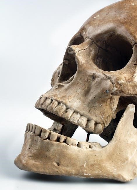
The facial skeleton, forming the anterior aspect of the skull, consists of fourteen bones providing structural support and defining facial features. Detailed PDF guides illustrate these: the paired maxillae forming the upper jaw, the palatine bones contributing to the hard palate, the zygomatic bones creating the cheekbones, and the paired nasal bones forming the bridge of the nose.
Additionally, the lacrimal bones contribute to the medial orbital wall, while the inferior nasal conchae increase the nasal cavity’s surface area. The vomer forms the inferior part of the nasal septum, and the mandible, the only movable facial bone, constitutes the lower jaw. Anatomy PDF resources highlight the foramina within these bones, facilitating passage for nerves and vessels.
Studying these bones, using textbooks and atlases, is crucial for understanding facial aesthetics and surgical approaches.
Hyoid Bone
The hyoid bone, a unique horseshoe-shaped structure in the anterior neck, doesn’t articulate with any other bone, instead suspended by ligaments and muscles. PDF anatomical resources detail its central body and paired greater horns (cornua), along with lesser horns, serving as attachment points for numerous muscles involved in swallowing and speech.
Its position inferior to the mandible is clearly illustrated in head and neck anatomy atlases. The hyoid’s stability relies on surrounding musculature, including the suprahyoid and infrahyoid groups. Understanding its relationships, often found in detailed textbooks, is vital for comprehending laryngeal function and airway management.
PDF guides emphasize its clinical significance in assessing airway obstruction and diagnosing related pathologies.
Muscles of the Head and Neck

Detailed PDF guides and textbooks illustrate the complex musculature of the head and neck, categorized by function – expression, mastication, and neck movement.

These resources aid in visualizing origins, insertions, and actions.
Muscles of Facial Expression
Facial expression muscles, intricately detailed in head and neck anatomy PDF resources and textbooks, are unique as they insert into the skin, enabling a diverse range of emotions and non-verbal communication.
These muscles, absent from bone attachment, are supplied by the facial nerve (VII), and are grouped around the eyes (orbicularis oculi), nose (nasalis), mouth (orbicularis oris, buccinator), and cheeks.
Understanding their individual actions – raising eyebrows, wrinkling the nose, smiling, or pursing the lips – is vital for clinicians, particularly in fields like plastic surgery and neurology.
PDF atlases provide clear visualizations of these superficial muscles, aiding in identifying their origins and insertions, while comprehensive textbooks delve into their innervation and functional interplay.
Mastery of this anatomy is essential for interpreting facial cues and diagnosing conditions affecting facial nerve function.
Muscles of Mastication
Muscles of mastication, crucial for chewing, are thoroughly illustrated in head and neck anatomy PDF guides and detailed textbooks like Netter’s, emphasizing their powerful function and complex interplay.
These include the masseter, temporalis, medial and lateral pterygoids, all innervated by the mandibular branch of the trigeminal nerve (V3). Each muscle contributes uniquely to jaw movements – elevation, protrusion, retraction, and lateral excursion.
The masseter and temporalis primarily elevate the mandible, while the pterygoids facilitate side-to-side and protrusive movements.
PDF atlases offer detailed views of muscle origins, insertions, and fiber directions, aiding in understanding their mechanical advantage.
Clinical relevance includes temporomandibular joint (TMJ) disorders, where dysfunction of these muscles is a key factor, necessitating precise anatomical knowledge.
Neck Muscles (Sternocleidomastoid, Trapezius)
Neck muscles, prominently the sternocleidomastoid (SCM) and trapezius, are extensively detailed in head and neck anatomy PDF resources and textbooks, showcasing their significant roles in head and shoulder movement.
The SCM, innervated by cranial nerve XI (accessory) and cervical nerves C2-C3, flexes, rotates, and laterally flexes the head. Its absence can indicate neurological issues.
The trapezius, also innervated by cranial nerve XI and cervical nerves C3-C4, elevates, depresses, retracts, and rotates the scapula, influencing neck extension and shoulder stability.
PDF atlases provide clear visualizations of muscle attachments and actions, crucial for understanding biomechanics.
Clinical relevance includes torticollis (wryneck) affecting the SCM and trapezius-related pain syndromes, demanding accurate anatomical comprehension for diagnosis and treatment.
Neuroanatomy of the Head and Neck
Neuroanatomy of the head and neck, detailed in PDF guides, focuses on cranial nerves and plexuses, essential for function and clinical understanding.
These resources illustrate pathways for sensation, motor control, and autonomic regulation.

Cranial Nerves
Cranial nerves, meticulously detailed in head and neck anatomy PDF resources, are twelve paired nerves emerging directly from the brain, responsible for diverse sensory and motor functions within the head and neck region;
These nerves—Olfactory (I), Optic (II), Oculomotor (III), Trochlear (IV), Trigeminal (V), Abducens (VI), Facial (VII), Vestibulocochlear (VIII), Glossopharyngeal (IX), Vagus (X), Accessory (XI), and Hypoglossal (XII)—innervate structures like the eyes, ears, nose, mouth, and muscles of facial expression and mastication.
PDF atlases and textbooks, such as Netter’s, provide comprehensive illustrations of nerve origins, pathways, and target structures, crucial for understanding neurological examinations and diagnosing related pathologies. Understanding their specific functions—sensory, motor, or mixed—is paramount for clinical application, and detailed PDF guides aid in mastering this complex neuroanatomy.
Cervical Plexus
The cervical plexus, thoroughly illustrated in head and neck anatomy PDF materials, is a network of spinal nerves originating from cervical vertebrae C1-C5, providing innervation to the posterior scalp, neck, and shoulders.
Key branches include the greater occipital nerve (sensory to the posterior scalp), lesser occipital nerve (sensory to the ear and lateral neck), and transverse cervical nerve (innervates the sternocleidomastoid and trapezius muscles).
PDF resources and textbooks like Gray’s Anatomy for Students detail the plexus’s formation and branching patterns, essential for understanding referred pain patterns and clinical presentations. Mastering the cervical plexus via detailed PDF guides is vital for diagnosing and treating conditions affecting the neck and upper limbs, offering a clear understanding of its anatomical relationships.
Brachial Plexus (Relevant portions)
The brachial plexus, often detailed in comprehensive head and neck anatomy PDF resources, extends from the cervical plexus (C5-T1) and innervates the upper limb; its relevant portions impact the neck’s lateral aspects.
Key branches like the suprascapular nerve (innervates rotator cuff muscles) and the long thoracic nerve (serratus anterior) can present with referred pain to the neck. PDF atlases and textbooks emphasize understanding these connections.
Detailed head and neck anatomy PDF guides illustrate how injuries to these nerves manifest as neck pain or weakness. Studying these portions is crucial for clinicians, as nerve compression or damage can cause radiating pain patterns. Mastering these anatomical relationships through visual PDF aids is essential for accurate diagnosis and treatment planning.

Vascular Anatomy of the Head and Neck
Detailed head and neck anatomy PDF resources illustrate the carotid and vertebral arteries, vital for cerebral blood flow, alongside venous drainage pathways.
Understanding these vascular networks, often visualized in textbooks, is crucial for surgical and diagnostic procedures.
Arteries of the Head and Neck (Carotid, Vertebral)
The carotid and vertebral arteries are paramount in supplying the head and neck with oxygenated blood, and their detailed anatomy is readily available in comprehensive head and neck anatomy PDF resources.
The common carotid artery bifurcates into the internal and external carotid arteries, each serving distinct regions. The internal carotid supplies the brain, while the external carotid nourishes the face, scalp, and neck structures.
Conversely, the vertebral arteries originate from the subclavian arteries and ascend through the transverse foramina of the cervical vertebrae, ultimately forming the basilar artery within the skull.
PDF atlases and textbooks, like Netter’s, meticulously depict these arterial pathways, their branches, and anastomoses, crucial for understanding potential clinical implications such as stroke or vascular injury.
Visualizing these complex networks through detailed illustrations aids in grasping their spatial relationships and functional significance.
Veins of the Head and Neck
Venous drainage of the head and neck is complex, involving numerous veins that ultimately converge into the internal jugular and external jugular veins, comprehensively illustrated in head and neck anatomy PDF guides.
The internal jugular vein, a major vessel, receives blood from the brain, face, and neck, while the external jugular vein drains superficial structures. These veins often accompany arteries, forming venous plexuses.

PDF resources, including detailed anatomical charts from textbooks like Gray’s Anatomy for Students, highlight the intricate network of smaller veins, such as the facial, temporal, and retromandibular veins.
Understanding venous anatomy is vital due to its clinical relevance in conditions like venous sinus thrombosis or facial swelling. Detailed PDF atlases aid in visualizing these pathways.
Accurate knowledge of venous drainage is crucial for surgical procedures and diagnosing various pathologies.
Regional Anatomy: Head
Detailed PDF resources illustrate the head’s regional anatomy, covering scalp layers, calvarium foramina, and underlying structures for comprehensive understanding and clinical correlation.
These guides aid in visualizing the complex relationships within the cranial region.
Scalp Layers
The scalp, a multi-layered structure covering the cranium, is meticulously detailed in head and neck anatomy PDF resources and textbooks. These layers, from superficial to deep, are commonly remembered using the mnemonic “SCALP”.
Skin, containing hair follicles and sebaceous glands, forms the outermost layer. Beneath lies the Connective Tissue layer, a dense, richly vascularized area. The Aponeurosis, or galea aponeurotica, is a tough, fibrous sheet connecting the frontalis and occipitalis muscles.
Deep to the aponeurosis is the Loose Connective Tissue layer, allowing movement of the scalp. Finally, the Pericranium, the periosteum of the skull, adheres directly to the bone. Understanding these layers, often visualized in anatomical PDFs, is crucial due to the scalp’s significant vascular supply and potential for extensive bleeding.
Calvarium and its Foramina
The calvarium, or cranial vault, forms the superior portion of the skull, extensively illustrated in head and neck anatomy PDF guides and detailed textbooks. It’s composed of the frontal, parietal, and occipital bones, protecting the brain.
Numerous foramina perforate the calvarium, serving as pathways for nerves and blood vessels. The foramen magnum, at the base of the occipital bone, transmits the spinal cord. The temporal foramina allow passage of the middle meningeal artery and nerve.
Smaller foramina, like those for emissary veins, connect the cranial and extracranial venous systems. Precise identification of these openings, often aided by PDF anatomical charts, is vital for understanding neurovascular pathways and potential clinical implications, such as intracranial hemorrhage.

Regional Anatomy: Neck
Neck anatomy, detailed in head and neck anatomy PDF resources, involves fascial layers and triangles—anterior and posterior—housing vital structures for study.
Understanding these regions, aided by textbooks, is crucial for clinical applications and comprehensive anatomical knowledge.
Fascial Layers of the Neck
The neck’s fascial layers, meticulously illustrated in head and neck anatomy PDF resources, are critical for understanding regional organization and spread of infection.
Superficial fascia contains cutaneous nerves, vessels, and fat; the deep fascia is subdivided into investing, pretracheal, prevertebral, and alar layers.
The investing layer envelops the sternocleidomastoid and trapezius muscles, while the pretracheal layer covers the trachea and thyroid gland.
Prevertebral fascia lies deep to the pretracheal layer, and the alar fascia attaches to the cranial base, aiding in compartmentalization.
These layers, detailed in anatomical textbooks, provide pathways for vessels and nerves, and define the boundaries of the neck’s triangles.
Studying these layers via PDF atlases enhances comprehension of surgical approaches and clinical presentations related to neck pathology.
Anterior and Posterior Triangles of the Neck
The neck is clinically divided into anterior and posterior triangles, a concept thoroughly detailed in head and neck anatomy PDF guides and comprehensive textbooks.
The anterior triangle, bounded by the midline, sternocleidomastoid, and mandible, contains muscles like the sternohyoid and omohyoid, and structures like the carotid sheath.
The posterior triangle, defined by the sternocleidomastoid, trapezius, and clavicle, houses the brachial plexus, subclavian artery, and several nerves.
Understanding these triangles, often visualized in anatomical PDF atlases, is crucial for identifying anatomical landmarks and potential surgical pathways.
Clinical relevance includes assessing for lymph node enlargement and understanding the spread of infection within these defined spaces.
Detailed study, utilizing resources like Netter’s, facilitates accurate diagnosis and treatment planning for neck pathologies.

Resources for Head and Neck Anatomy Study
Essential study aids include Netter’s and Gray’s textbooks, alongside readily available head and neck anatomy PDF atlases for detailed visualization.
Online resources further supplement learning, offering interactive models and comprehensive anatomical charts.
Netter’s Head and Neck Anatomy
Netter’s Head and Neck Anatomy stands as a cornerstone resource for students and practitioners alike, renowned for its beautifully illustrated and clinically focused approach. This textbook excels in presenting complex anatomical structures with exceptional clarity, utilizing vibrant illustrations that significantly enhance understanding.
Many students supplement the physical textbook with a head and neck anatomy PDF version for convenient study on various devices. The PDF format allows for easy searching, annotation, and portability, making it ideal for quick reference during clinical rotations or exam preparation.
The book meticulously covers osteology, myology, neuroanatomy, and vascular anatomy, providing a comprehensive overview of the region. Its clinical correlations highlight the relevance of anatomical knowledge to real-world medical scenarios, solidifying learning and promoting practical application. It’s a highly recommended resource for mastering the intricacies of the head and neck.
Gray’s Anatomy for Students
Gray’s Anatomy for Students offers a streamlined and accessible approach to the vast subject of anatomical study, specifically tailored for medical, dental, and allied health students. While the comprehensive print version remains a classic, a head and neck anatomy PDF version is frequently utilized for its convenience and searchability.
This edition focuses on clinically relevant anatomy, emphasizing the structures and concepts most important for understanding patient care. It integrates clinical imaging, surface anatomy, and histological images to provide a holistic learning experience.
The PDF format allows students to easily navigate the text, highlight key information, and access supplementary materials. It complements the detailed illustrations with concise explanations, making it an effective tool for both self-study and exam preparation. It’s a valuable resource alongside other textbooks and anatomical atlases.
Online Anatomy Atlases and PDFs
Numerous online resources provide head and neck anatomy PDF materials, supplementing traditional textbooks like Netter’s and Gray’s. These digital atlases offer interactive 3D models, labeled diagrams, and detailed illustrations, enhancing visualization and comprehension. Many universities and medical institutions offer free access to their anatomy resources, including downloadable PDF study guides.
Websites dedicated to anatomy often feature quizzes and self-assessment tools to reinforce learning. Caution should be exercised when downloading PDFs from unfamiliar sources to ensure accuracy and avoid copyright infringement.
Reputable platforms provide high-quality, peer-reviewed content, making them invaluable for students and healthcare professionals seeking a convenient and accessible way to study the complex anatomy of the head and neck.